Apr 09, 2024PRESS RELEASE
Visualization of the True Distribution State of Trace Uranium using Superconducting Technology
~ Expectations for Understanding the Migration Behavior of Various Trace Elements in the Environment ~
Keyword:INFORMATION
OBJECTIVE.
The understanding of the migration behavior of uranium (U), used as fuel for nuclear power generation, in the environment is important for the safety assessment during the disposal of radioactive waste. In order to accurately grasp the migration behavior of U in the environment, there was a need for a new analytical technique that can detect only the signal of trace amounts of U among the signals of many elements in a sample.
Associate Professor Shinya Yamada of the Department of Science at Rikkyo University, Professor Yoshio Takahashi of the Graduate School of Science at The University of Tokyo, Researcher Takumi Yomogida of the Japan Atomic Energy Agency, Term-appointed Researcher Tomoya Uruga of the Japan Synchrotron Radiation Research Institute (JASRI), Researcher Kiyofumi Nitta, and Chief Researcher Oki Sekizawa have conducted joint research with multiple research institutions that promote the use of a superconducting transition-edge sensor (Transition Edge Sensor; TES) (Note 1), which can detect signals of specific energies with high energy resolution. This time, at beamline BL37XU of the large-scale synchrotron radiation facility SPring-8 (Note 2), they have for the first time in the world applied TES as a detector for fluorescent XAFS (X-ray Absorption Spectroscopy) (Note 3) analysis using a microbeam X-ray, successfully grasping the distribution state of trace amounts of U in real environmental samples, which cannot be captured by conventional semiconductor detectors.
This research enables the analysis of ultra-trace elements in environmental samples with micrometer-sized spatial resolution, and by elucidating the mechanism of element migration behavior at the atomic and molecular scale, it is expected to expand to studies on the environmental migration behavior of various elements, not just U.
Furthermore, TES is being developed for various applications such as space X-ray observation, atomic and molecular physics, and nuclear physics, with ongoing research on device development and applicability. The results of this study, which succeeded in analyzing real environmental samples non-destructively, are widely expected to be applied to the non-destructive analysis of extraterrestrial samples obtained from future small celestial body sample return missions, as well as to the earth, environmental, extraterrestrial samples, and biological samples.
This research will be published online at 16:00 Japan time on April 9, 2024, by the Royal Society of Chemistry's journal 'Analyst'.
Research Background:
Uranium (U) is widely used as fuel for nuclear power generation around the world. There are two methods being considered for the disposal of spent nuclear fuel generated in this process: direct disposal without reprocessing, or indirect disposal where radioactive waste generated during reprocessing is disposed of. In either case, the radioactive waste is planned to be buried underground, and especially in the case of the former, research to understand the migration behavior of U in the subterranean environment is considered important. The solubility of U in water can vary greatly depending on its chemical state; thus, knowing the chemical and distribution state of U in environmental samples leads to the estimation of its migration behavior. Fluorescence X-ray absorption fine structure analysis using synchrotron radiation (fluorescent XAFS method) is a technique that, in principle, allows for the analysis of the chemical state (valence and bonding state) of any element by precisely measuring the energy of the fluorescence X-rays and scattered X-rays emitted from the sample. Furthermore, by focusing the X-rays irradiated on the sample to micrometer size, it is possible to examine the distribution and chemical state of elements with high spatial resolution. However, in environmental samples containing various elements, the energy resolution of the commonly used semiconductor detectors is insufficient, and the fluorescence X-rays from trace amounts of U can be obscured by the fluorescence X-rays of other elements (such as Rubidium (Rb)) that are abundant in the earth's crust, making it difficult to accurately grasp the distribution state and chemical species. The research group focused on a spectrometer called a superconducting transition-edge sensor (Transition Edge Sensor; TES), which has both high energy resolution and high detection efficiency, to challenge this issue.
Research Results:
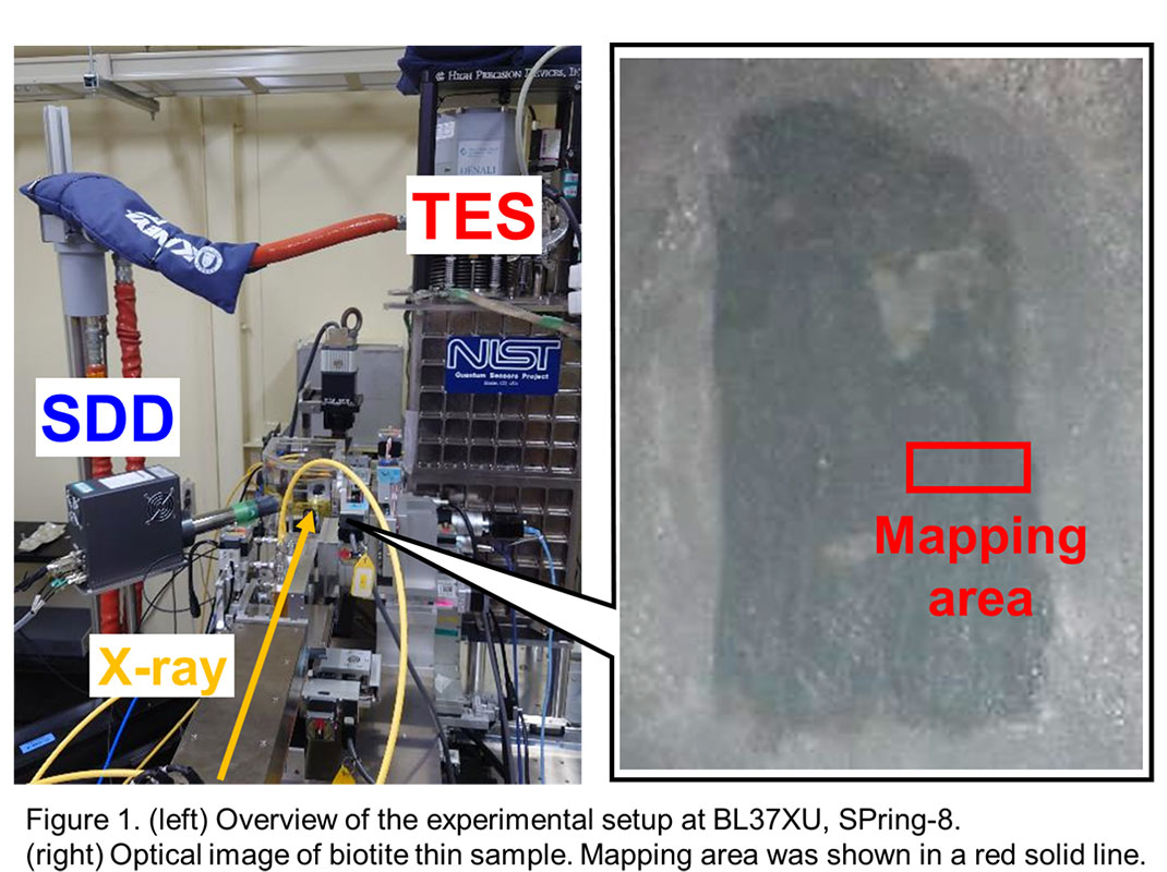
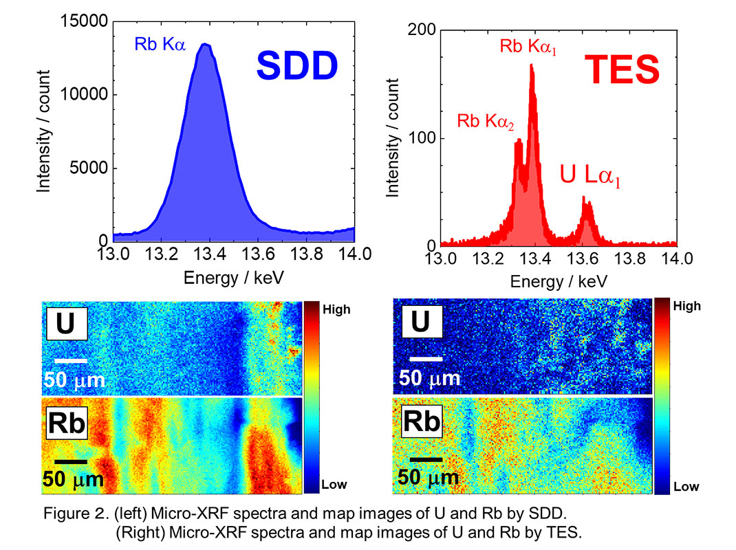
The research group conducted an operational demonstration of TES (made by NIST in the USA) brought to the SPring-8 beamline BL37XU, using both TES and a semiconductor detector (Silicon Drift Detector: SDD). Figure 1 shows the experimental setup at SPring-8 BL37XU and the appearance of the biotite sample collected from the environment used in this analysis. Figure 2 compares the mapping analysis results using microbeam X-rays with the SDD and TES in the area indicated on the right side of Figure 1. The conventional SDD could only observe the fluorescence X-ray peak of Rb, which is abundantly contained in biotite, and could not accurately detect the signal of trace amounts of U. Therefore, the distribution of U appeared similar to that of Rb, and it was not possible to obtain an accurate distribution state of U. On the other hand, the results of the analysis using TES are on the right side of Figure 2. From the measurement results of the fluorescence X-ray spectrum, it is clear that TES could separate and measure the fluorescence X-rays from trace amounts of U, which were impossible to extract with the SDD. Furthermore, the ability to accurately extract the U signal confirmed that the distribution of Rb and U is different. These results show that by using TES, it is possible to accurately understand the distribution state of trace amounts of U, which was difficult to analyze with conventional detectors. In addition, from the results of the XAFS measurements performed simultaneously, the analysis of the chemical state of U contained in biotite was successful, and it became clear that some of the U in biotite was reduced. This indicates that U has become less mobile in the strata as a result of being reduced and fixed in the biotite, shedding light on part of the mechanism by which biotite retains U.
Although this experiment focused on U and Rb in environmental samples, it was confirmed that TES has high energy resolution up to the high energy region of 17 keV. Therefore, it has been demonstrated that TES can be applied not only to U but also to the analysis of other elements that have fluorescence X-rays in the energy region up to 17 keV, and the application to various environmental samples is expected in the future.
“Application of transition-edge sensor for micro-X-ray fluorescence measurements and micro-X-ray absorption near edge structure spectroscopy: a case study of uranium speciation in biotite obtained from uranium mine”
T. Yomogida et al., Analyst. (2024), doi: 10.1039/D4AN00059E.
Future Developments:
Measurements that were previously impossible over a wide energy range with high-energy resolution, as well as a drastic reduction in measurement time, will now make it possible to significantly reduce sample damage. TES technology is evolving rapidly, and with advances in speed and increased effective area, applications in the analysis of the chemical states of ultra-trace elements in environmental samples, astrochemical samples, and biological samples are expected. In the future, by achieving higher energy resolution, further developments in advanced high-energy resolution fluorescence X-ray detection methods such as the X-ray Absorption Near Edge Structure method with high energy resolution fluorescence detection (HERFD-XANES method (Note 4) are also expected. We aim to continue advancing the maturity of this technology and develop TES-based research in fields that are broadly beneficial to society, such as basic sciences including space X-ray observation, atomic and molecular physics, and nuclear physics, as well as non-destructive analysis in future sample return missions and environmental science for the realization of a sustainable society.
Footnotes:
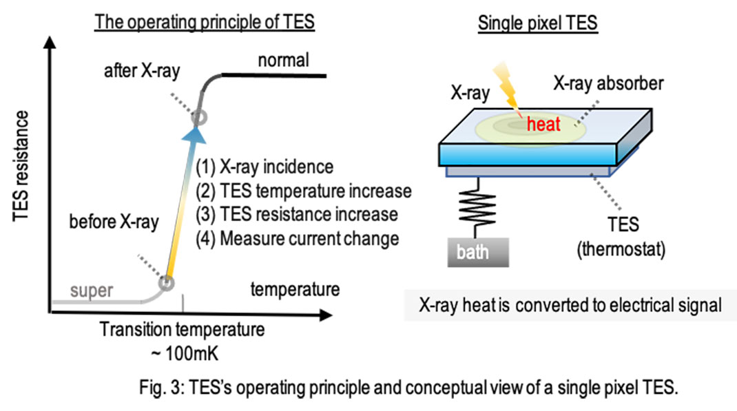
- (Note 1) A Transition Edge Sensor (TES) is a detector that utilizes the sharp resistance-temperature characteristic near the superconducting-normal phase transition point. As shown in Figure 3, when X-rays are absorbed by the detector's heat-absorbing part, the temperature of the TES increases, as does its resistance value. Detecting the change in current flowing through the circuit allows for the estimation of the energy of the incident X-rays. The significant change in resistance value at the phase transition point of the superconductor allows for accurate energy estimation.
- (Note 2) Located in Harima Science Park City, Hyogo Prefecture, this facility at the RIKEN Institute produces the world's highest-performance synchrotron radiation and is supported by the Japan Synchrotron Radiation Research Institute (JASRI). The name SPring-8 is derived from Super Photon ring-8 GeV (gigaelectronvolts). Synchrotron radiation is a highly directional and powerful electromagnetic wave generated when electrons, accelerated to speeds nearly equivalent to that of light, are bent by magnets. SPring-8 utilizes this synchrotron radiation for a wide range of research, from nanotechnology and biotechnology to industrial applications.
- (Note 3) Fluorescence detection X-ray Absorption Fine Structure (XAFS) is a method that analyzes the fine structure of X-ray absorption obtained by analyzing fluorescence X-rays of various wavelengths generated when X-rays are irradiated onto a substance. This allows for the investigation of the elements contained within the material, their types, and chemical states.
- (Note 4) High-Energy-Resolution Fluorescence Detected X-ray Absorption Near Edge Structure (HERFD-XANES) is a method within fluorescence XAFS for finely separating (spectroscopy with high energy resolution) specific wavelengths of fluorescence X-rays. Traditionally, the use of crystals for spectroscopy was necessary, which had the disadvantage of discarding much useful information. However, the use of a non-dispersive spectrometer like TES is considered to enable efficient measurements.
Research Support
This research was supported by JSPS KAKENHI (Grant Numbers: (18H05458, 19H01145, 19H01960, 19K15606, 19K21893, 19K23432, 20K20527, 20K15238, 20K14524, 21H00162, 21H03585, 21H05443, 21K18649, 21K18917, 22F21313, 22H00166, and 22K18277)) and projects at JASRI/SPring-8 and the Photon Factory (2022A0174, 2022A0180, 2021A1610, 2019A1523, 2019B1498, and 2020A0174)、Photon Factory (2022G126, 2020G670, 2020G081, and 2018S1-001)).
Contribution
T. Yomogida and Y. Takahashi designed the research and wrote the paper. T. Yomogida. performed the sample preparation, the synchrotron measurements, and analyses. T. Hashimoto, T. Okumura, S. Yamada, H. Tatsuno, H. Noda, R. Hayakawa, S. Okada, S. Takatori, T. Isobe, T. Hiraki, T. Sato, Y. Ichinohe, Y. Toyama, O. Sekizawa, K. Nitta and T. Uruga performed the synchrotron measurements, contributed to manuscript writing. Y. Kurhihara. and S. Fukushima contributed to the acquisition of biotite sample. Y. Kitatsuji contributed to manuscript writing. All the authors discussed the results and contributed to the manuscript.